How to Detect Host Cell Protein in Biologics?
Detecting host cell protein in biologics is not a simple matter of obtaining a low assay value. These process-related impurities can originate from the expression system, harvesting step, clarification stage, or purification train. Their composition also changes with process conditions. A single test may miss important proteins.
Peter Gagnon, a recognized protein-purification specialist, has stated, “The real challenge is not measuring HCPs, but understanding what the measurement means.” This principle remains practical. ELISA can provide sensitive, routine monitoring when its antibody coverage fits the production platform. It may still overlook unexpected or poorly immunogenic proteins. LC-MS can add broader identification and characterization. It can also introduce complex data interpretation.
Details matter.
A reliable host cell protein strategy begins with representative process samples. Early harvest material, intermediate pools, and final drug substance should be compared. Analysts should review dilution effects, matrix interference, recovery, precision, and assay suitability. Orthogonal methods can help confirm whether an apparent decrease reflects genuine clearance or merely reduced detectability.
Experience often reveals uncomfortable gaps. A validated assay is not automatically a complete assay. A low result is not automatically proof of low risk. Some decisions depend on process history, platform knowledge, impurity identity, and toxicological relevance. Method performance should therefore be assessed against the molecule and manufacturing process, not only against a standard curve.
This article explains how to detect host cell protein in biologics, select complementary technologies, and interpret results responsibly. It also examines common analytical weaknesses, because dependable conclusions require more than impressive numbers.
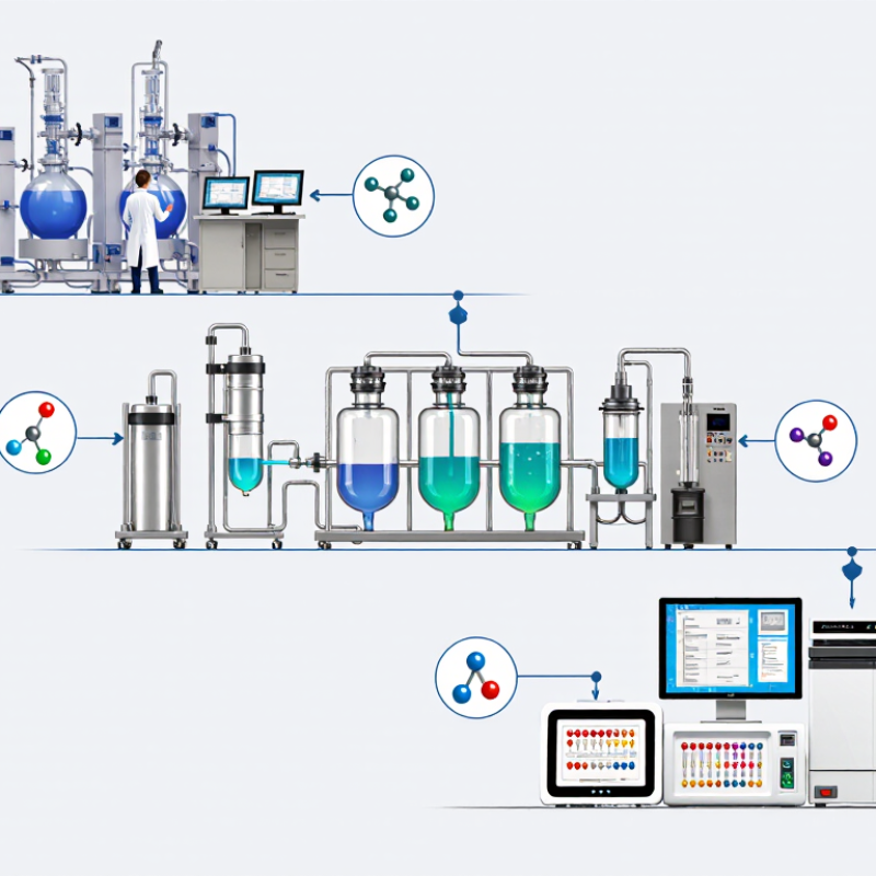
What Are Host Cell Proteins and Why Must They Be Detected?
Host cell proteins are unintended proteins left behind after a biologic is produced in living cells. The manufacturing cells create the therapeutic molecule, but they also release thousands of their own proteins. These residues may include enzymes, chaperones, membrane proteins, or fragments from damaged cells. They are not part of the intended medicine.
Why does detection matter? Even tiny amounts can affect safety, stability, or immune responses. Some proteins may trigger inflammation or reduce product quality over time. Others can interfere with purification or change during storage. The risk depends on the protein, its concentration, and the patient’s exposure. More impurity does not always mean greater danger. That assumption can fail.
In practice, laboratories often screen samples with sensitive immunoassays and investigate selected samples using mass spectrometry. The first method can measure broad HCP levels efficiently. The second can help identify individual proteins that routine testing may miss. Sample preparation is critical. Poor recovery can make a clean sample look safer than it is. No method is flawless. Therefore, analysts compare results across process stages, testing purified material, intermediate fractions, and stability samples. They also examine trends rather than relying on one number. A falling HCP level is encouraging, but it does not prove that every relevant protein has disappeared. Careful method validation, qualified reference materials, and documented laboratory experience support reliable decisions.
How to Prepare Biologic Samples for HCP Analysis
Reliable host cell protein analysis begins with a representative sample. Collect material from the correct process stage, using clean containers and documented handling conditions. Mix gently to prevent settling, foaming, or protein aggregation. Record the sample concentration, buffer composition, storage time, and number of freeze-thaw cycles. Small details matter.
Clarify visibly turbid samples by centrifugation when the method permits. Avoid unnecessary filtration because filters may remove proteins or introduce extractable contaminants. Dilute highly concentrated biologics within the assay’s validated range. Use the same diluent for standards, controls, and test samples whenever possible. Prepare separate aliquots to reduce repeated freezing. Keep samples cold, but do not assume colder is always better.
Include blank matrix, negative process controls, and a fortified recovery sample. These controls can reveal dilution effects, interference, or sample loss. A spike recovery result may look acceptable while certain HCP classes remain poorly detected. That limitation deserves attention. Analysts should compare dilution linearity across more than one level and investigate unexpected signal changes. If the sample contains antibodies, lipids, or formulation excipients, test for assay interference before interpreting low HCP values. A carefully prepared sample supports credible results, but preparation alone cannot prove complete HCP coverage. Method suitability, orthogonal evidence, and documented deviations remain essential.
| Biologic Sample Type | Typical Sample Matrix | Recommended Sample Preparation | Typical Starting Dilution | Key Parameters to Control | Main HCP Analysis Considerations |
|---|---|---|---|---|---|
| Purified monoclonal antibody | Protein solution, commonly containing salts, buffering agents, and stabilizers | Mix thoroughly. Dilute with a validated assay diluent. Clarify visible particles by low-speed centrifugation or filtration when appropriate. Avoid repeated freeze–thaw cycles. | Often 1:10 to 1:1,000, depending on product concentration and assay range | Protein concentration, pH, ionic strength, viscosity, surfactant level, and matrix background | High product concentration may cause signal suppression or nonspecific interference; dilutional linearity should be demonstrated. |
| Recombinant protein | Purified protein in a formulation or process intermediate | Dilute into assay diluent. If the sample contains precipitate, centrifuge and test the clarified fraction; document whether particulate material was removed. | Commonly 1:5 to 1:500 | Aggregation, precipitation, reducing agents, detergents, and high salt concentrations | Aggregates can adsorb HCP or affect immunoassay recovery. A qualified sample-treatment step may be required. |
| Viral vector or virus-like particle | Vector-containing solution with capsid, nucleic acid, salts, and process-related impurities | Use gentle mixing. Dilute carefully and avoid conditions that disrupt or precipitate the particle unless intentional lysis is part of the validated method. | Often 1:10 to 1:200 | Particle concentration, nuclease residues, detergents, pH, and sample turbidity | Matrix effects may differ between intact and disrupted particles. The preparation protocol should define whether total accessible HCP or soluble HCP is measured. |
| Plasma-derived or serum-containing product | Complex protein-rich biological matrix | Clarify by centrifugation if needed, dilute substantially, and assess nonspecific binding. Consider orthogonal confirmation using a mass-spectrometric method. | Frequently 1:100 to 1:10,000 | Endogenous proteins, lipids, hemolysis, turbidity, and high-abundance matrix components | Immunoassay selectivity can be limited by the complex matrix; spike recovery and parallelism are especially important. |
| Cell-culture harvest | Unpurified harvest containing cells, cell debris, media components, and soluble HCP | Clarify by validated centrifugation and/or depth filtration. Record clarification conditions and analyze the clarified supernatant. | Commonly 1:100 to 1:10,000 | Cell density, debris load, viscosity, storage time, and protease activity | The measured HCP profile may change during clarification because cell-associated proteins and insoluble material are removed. |
| Chromatography pool | Partially purified process intermediate containing buffer and elution components | Mix thoroughly and dilute into a compatible assay diluent. If the buffer is strongly acidic or basic, neutralize only when the validated procedure permits it. | Often 1:10 to 1:1,000 | pH extremes, conductivity, chaotropes, detergents, and residual cleaning or elution reagents | Buffer components can alter antibody–antigen binding and produce nonparallel dilution curves. |
| Final drug product | Filled formulation containing the active biologic and excipients | Invert or gently mix. Reconstitute lyophilized material with the specified diluent. Analyze within the established hold-time window. | Frequently 1:10 to 1:1,000 | Excipients, surfactants, sugar concentration, container interaction, and reconstitution time | Formulation components may cause assay interference or alter HCP recovery; matrix-matched controls are recommended. |
| Stability or stressed sample | Material exposed to defined temperature, light, agitation, or freeze–thaw conditions | Use the same preparation procedure as the unstressed control. Record exposure history and inspect for visible particles or turbidity before testing. | Use the dilution that provides acceptable parallelism and recovery | Aggregation, degradation, precipitation, adsorption, and sample hold time | Stress can change HCP accessibility and product matrix behavior, so results should be compared with appropriate controls. |
| Sample-Preparation Checkpoint | Practical Guidance | Why It Matters for HCP Results |
|---|---|---|
| Homogenization | Mix gently but thoroughly; avoid foaming. | Incomplete mixing can produce nonrepresentative aliquots and poor repeatability. |
| Clarification | Use centrifugation or filtration only when compatible with the analytical objective. | Removing particles may also remove particle-associated HCP and change the reported result. |
| Dilution linearity | Evaluate multiple dilutions across the assay range. | Parallel dilution responses help identify matrix interference and dilution-dependent bias. |
| Spike recovery | Add a known HCP control to representative samples and calculate recovery. | Recovery demonstrates whether the product matrix suppresses or enhances assay signal. |
| Freeze–thaw control | Prepare single-use aliquots whenever possible. | Repeated cycles may cause aggregation, adsorption, degradation, or changes in HCP accessibility. |
| Orthogonal confirmation | Use a complementary method such as targeted or untargeted mass spectrometry when justified. | Different methods provide complementary information about HCP identity, coverage, and immunoreactivity. |
Which Methods Are Used to Detect Host Cell Proteins?
How to Detect Host Cell Protein in Biologics?
Which Methods Are Used to Detect Host Cell Proteins?
Host cell protein detection protects biologic quality and patient safety. These residual proteins come from the cells used during production. A validated ELISA is often the routine screening method. It uses antibodies against a broad range of possible host cell proteins. Results can be sensitive, but antibody coverage is never perfect.
LC-MS/MS provides deeper identification and can detect proteins missed by immunoassays. It separates peptides, measures their mass, and compares signal patterns with reference databases. Two-dimensional electrophoresis can show protein spots with different abundance levels. However, it may miss low-level or poorly soluble proteins. Western blotting can support investigation when a specific protein is suspected. No single method answers every question.
Tips: Use orthogonal methods when risk is high. Compare samples from different purification stages. Check assay recovery, dilution linearity, and matrix effects. Keep sample handling consistent; small temperature changes can affect results. Experienced analysts should review unexpected peaks manually. Automated reports are useful, but they can simplify difficult biology. Some proteins remain undetected because the test design is incomplete. That limitation deserves honest discussion.
How to Quantify and Interpret HCP Results
Detecting host cell protein (HCP) is not only an assay exercise. It is a measurement problem requiring controlled evidence. During development, I review sample history, purification steps, and assay suitability together. An HCP ELISA usually reports concentration through a calibrated standard curve. The curve should show acceptable fit, precision, and defined lower limits of quantification. Prepare samples at suitable dilutions. Confirm dilutional linearity, spike recovery, and replicate agreement. Keep records of plate effects, analyst actions, and unexpected results.
Do not treat one HCP value as the complete risk picture. Compare results across process stages and manufacturing lots. A rising value after purification may indicate process variation, sample interference, or altered protein recognition. It needs investigation. Report results with units, dilution factors, acceptance criteria, and uncertainty where possible. Very low readings may reflect genuine clearance. They may also approach the assay’s quantification limit. That distinction matters.
Orthogonal testing can strengthen interpretation. Mass spectrometry may reveal HCP species that an antibody-based assay detects weakly or misses. However, it also requires careful digestion, identification, and quantitative controls. A result without suitable controls is fragile. Sometimes the data look clean, but the evidence is incomplete. I would revisit recovery experiments, matrix effects, and reagent specificity before making a firm conclusion. Experienced reviewers should examine trends, not just pass-or-fail numbers. Reliable HCP control depends on method performance, process knowledge, and honest recognition of uncertainty.
How to Detect Host Cell Protein in Biologics?
How to Quantify and Interpret HCP Results
What the chart shows: Residual host cell protein (HCP) concentration is reported as nanograms per milligram of product protein (ng/mg). The representative purification profile shows progressive HCP clearance from harvest through the final drug substance.
How to interpret the result: HCP is commonly quantified with a validated immunoassay, such as a platform or product-specific ELISA. Results should be assessed together with assay accuracy, precision, dilutional linearity, spike recovery, specificity, and the validated lower limit of quantification (LLOQ).
Important: There is no universal HCP acceptance limit for every biologic. The final specification is product- and process-specific and should be justified using process capability, toxicological risk assessment, clinical experience, and regulatory expectations.
How to Validate HCP Testing and Investigate Unexpected Findings
Host cell protein testing needs more than a passing assay result. It requires evidence that the method fits the product matrix. Validation should examine specificity, accuracy, precision, linearity, sensitivity, and dilutional parallelism. Spike recovery can reveal whether the sample suppresses or exaggerates detection. A low signal is not automatically good news.
In practice, unexpected HCP results often begin with simple questions. Was the sample mixed thoroughly? Did storage time exceed the approved window? Were controls within range? Review analyst records, plate maps, reagent preparation, and instrument performance. Compare the result with historical process trends, not one isolated batch. If the finding remains unusual, test a retained sample using an orthogonal approach. This may expose assay interference or an incomplete impurity profile. Sometimes, the investigation itself is imperfect. That limitation should be documented, not hidden.
Tips: Build a matrix-specific validation plan. Include stressed and representative samples. Define acceptance criteria before testing. Investigate both unusually high and unexpectedly low results. Check dilution behavior across multiple levels. Use independent review for atypical data. Keep raw observations, including failed runs, because they may explain later trends. Ask whether the assay measures the risk relevant to the process. A technically valid method may still miss important proteins.
Related Posts
-
Why Are Host Cell Proteins Important in Bioprocessing?
-

Top 10 Host Cell Protein ELISA Applications You Should Know?
-
10 Essential Tips for Understanding Host Cell Proteins in Biotechnology
-

Best Host Cell Protein Products for Global Buyers?
-

2026 Top Mycoplasma Detection Techniques for Global Buyers?
-

Top Recombinant Protein Suppliers for Global Buyers?